
 Copy to clipboard
Nathan Skelley, M.D., is motivated by the continuous pursuit of performing increasingly complex sports medicine procedures through smaller incisions. He believes that orthopedic surgeons must constantly tap available resources to stay informed about the latest surgical technologies, buy into data-driven care to determine ways to improve patient outcomes and be willing to learn and adapt to the latest trends.
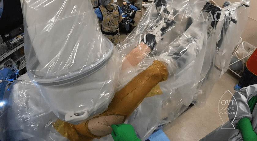
Dr. Skelley, a fellowship-trained sports medicine surgeon at Sanford Health and the University of South Dakota School of Medicine, actively pursues innovative solutions for treating a variety of musculoskeletal conditions. He and his colleagues won an Orthopedic Video Theater Award at this year’s AAOS Annual Meeting for demonstrating an alternative arthroscopic approach that preserves hip capsule anatomy.
Here he discusses the award-winning hip arthroscopy technique, the increasing role of biologics in advancing patient care and an out-of-this-world application for the 3D-printed external fixator he helped develop.
What current trends in sports medicine are capturing your attention and interest?
Dr. Skelley: I’m particularly excited by recent advancements in enhanced visualization systems that provide clearer images during arthroscopic procedures, as well as the emergence of biologics and devices that complement mechanical repairs by facilitating tissue healing and ligament reconstruction. Virtual reality (VR) and augmented reality (AR) are transforming surgical training by offering a more immersive and realistic learning experience for the next generation of surgeons. I think that’s a significant development.
Numerous orthopedic companies are investing in solutions aimed at preserving joint health and delaying or eliminating the need for joint replacement surgery. What developments in this space show promise?
Dr. Skelley: Biologics are becoming increasingly relevant in sports medicine. Arthrex has made significant strides with platelet-rich plasma (PRP) and bone marrow aspirate concentrate (BMAC) systems for hip core decompression surgery. Additionally, MTF Biologics has introduced CartiMax, a revolutionary solution for cartilage defects in the knee.
This technology is particularly exciting as it simplifies the tedious process of suturing in cartilage grafts. CartiMax offers a more straightforward approach — it’s a putty that can be applied directly into defects. Histologic studies have demonstrated its ability to promote the growth of hyaline cartilage, which is the ultimate goal.
CartiMax currently needs to be applied through open incisions, but there’s hope that surgeons will eventually transition to applying it during arthroscopic surgeries. This advancement holds immense promise for the field and hopefully, we’ll witness its implementation in minimally invasive procedures in the next few years.
CONMED’s BioBrace is also an intriguing application. It contains bovine collagen reinforced with PLLA and functions as a bioinductive implant. Essentially, it offers mechanical strength and biological healing potential. Presented results involving the implant are promising and highlight the additional value of using biologics to augment mechanical repairs.
Enabling technology is gaining momentum in joint replacement and spine. Do you see the same level of interest in sports medicine?
Dr. Skelley: Not to the same degree, but some of the advanced imaging and navigation technologies that are commonly associated with robotics have proven useful in sports medicine applications. Stryker’s HipMap, for instance, has revolutionized my approach to hip arthroscopy. The system employs CT scans to create 3D models of bone impingement locations and alignment of the hip joint. This provides a wealth of information that guides my approach during surgery.
Before having access to the technology, I often felt like I was operating blindly — relying solely on x-rays, CT scans and dynamic examinations in the operating room to detect impingements and other pathology within the joint. Now, HipMap lays out that critical information before I enter the operating room. Our research demonstrated a strong correlation between the outcomes predicted by the scans and what we observed during surgery. The tool has become an integral part of my practice for all hip arthroscopy patients.
How is your capsule-preserving hip arthroscopy technique performed, and why is it better than the standard surgical approach?
Dr. Skelley: The traditional method of performing hip arthroscopy involves making large capsulotomies, which compromise the integrity of the robust ligaments surrounding the hip joint. This can lead to hip instability and trauma to the joint’s tissue post-surgery. Additionally, repairing the capsule at the end of the procedure is tedious and time-consuming, often adding up to 30 minutes to the surgery.
In contrast, our approach utilizes a portal approach through small incisions, starting in the peripheral compartment with an intact capsule. This minimally invasive surgery allows the arthroscopic fluid to fill the capsule like a balloon, providing a clear view and workspace within the joint. Starting in the peripheral compartment provides direct visualization of the femoral head, reducing the risk of damaging the labrum or cartilage during the procedure.
This approach promises safer surgery, faster recovery time, less postoperative pain and more effective outcomes for patients with hip arthritis.
Personalized patient care is driving much of the innovation in orthopedics today. How do you see that movement impacting your specialty?
Dr. Skelley: I used to follow a standardized approach to procedures, moving from one step to the next in a predictable manner. However, now I find myself adjusting my techniques based on the needs of individual patients. For example, HipMap might inform me that I need to position the portal higher to access a specific location due to known impingement issues or allocate additional operative time to address a pincer lesion identified on the MRI.
Although we haven’t conducted formal research on the benefits of personalized surgical approaches, I have observed anecdotally that patients tend to have better outcomes when approaches are tailored to address specific pathology present in the hip. This underscores the importance of compiling patient data and conducting a retrospective review to further investigate these observations.
You were actively involved in the development of a 3D-printed external fixator that could revolutionize the future of fracture repair. How have those efforts progressed?
Dr. Skelley: About two years ago, the University of South Dakota, in collaboration with the American Orthopaedic Society for Sports Medicine, secured funding for research focusing on the rural applications of 3D printing in orthopedics. I was inspired by the possibility of treating the emergent need for external fixators to treat fractures in injured civilians and soldiers on both sides of the Russian/Ukraine war.
Our research team embarked on the development of an external fixator device, the design of which has been published along with a systematic review of similar initiatives in the field. Recently, we have completed mechanical testing on the device and compared its performance to industry standards. Surprisingly, our findings revealed that the 3D-printed, low-cost external fixator was comparable in many of the mechanical tests that we conducted. This represents an exciting advancement in fracture repair, demonstrating the promising potential of 3D printing technology in orthopedics.
The application of 3D printing in this context essentially enables a print-on-demand model, eliminating the need to ship external fixators to specific locations or have them readily available. This has significant implications for remote research installations, forward-operating military units and long-duration spaceflight.
Nathan Skelley, M.D., is motivated by the continuous pursuit of performing increasingly complex sports medicine procedures through smaller incisions. He believes that orthopedic surgeons must constantly tap available resources to stay informed about the latest surgical technologies, buy into data-driven care to determine ways to improve...
Nathan Skelley, M.D., is motivated by the continuous pursuit of performing increasingly complex sports medicine procedures through smaller incisions. He believes that orthopedic surgeons must constantly tap available resources to stay informed about the latest surgical technologies, buy into data-driven care to determine ways to improve patient outcomes and be willing to learn and adapt to the latest trends.
Dr. Skelley, a fellowship-trained sports medicine surgeon at Sanford Health and the University of South Dakota School of Medicine, actively pursues innovative solutions for treating a variety of musculoskeletal conditions. He and his colleagues won an Orthopedic Video Theater Award at this year’s AAOS Annual Meeting for demonstrating an alternative arthroscopic approach that preserves hip capsule anatomy.
Here he discusses the award-winning hip arthroscopy technique, the increasing role of biologics in advancing patient care and an out-of-this-world application for the 3D-printed external fixator he helped develop.
What current trends in sports medicine are capturing your attention and interest?
Dr. Skelley: I’m particularly excited by recent advancements in enhanced visualization systems that provide clearer images during arthroscopic procedures, as well as the emergence of biologics and devices that complement mechanical repairs by facilitating tissue healing and ligament reconstruction. Virtual reality (VR) and augmented reality (AR) are transforming surgical training by offering a more immersive and realistic learning experience for the next generation of surgeons. I think that’s a significant development.
Numerous orthopedic companies are investing in solutions aimed at preserving joint health and delaying or eliminating the need for joint replacement surgery. What developments in this space show promise?
Dr. Skelley: Biologics are becoming increasingly relevant in sports medicine. Arthrex has made significant strides with platelet-rich plasma (PRP) and bone marrow aspirate concentrate (BMAC) systems for hip core decompression surgery. Additionally, MTF Biologics has introduced CartiMax, a revolutionary solution for cartilage defects in the knee.
This technology is particularly exciting as it simplifies the tedious process of suturing in cartilage grafts. CartiMax offers a more straightforward approach — it’s a putty that can be applied directly into defects. Histologic studies have demonstrated its ability to promote the growth of hyaline cartilage, which is the ultimate goal.
CartiMax currently needs to be applied through open incisions, but there’s hope that surgeons will eventually transition to applying it during arthroscopic surgeries. This advancement holds immense promise for the field and hopefully, we’ll witness its implementation in minimally invasive procedures in the next few years.
CONMED’s BioBrace is also an intriguing application. It contains bovine collagen reinforced with PLLA and functions as a bioinductive implant. Essentially, it offers mechanical strength and biological healing potential. Presented results involving the implant are promising and highlight the additional value of using biologics to augment mechanical repairs.
Enabling technology is gaining momentum in joint replacement and spine. Do you see the same level of interest in sports medicine?
Dr. Skelley: Not to the same degree, but some of the advanced imaging and navigation technologies that are commonly associated with robotics have proven useful in sports medicine applications. Stryker’s HipMap, for instance, has revolutionized my approach to hip arthroscopy. The system employs CT scans to create 3D models of bone impingement locations and alignment of the hip joint. This provides a wealth of information that guides my approach during surgery.
Before having access to the technology, I often felt like I was operating blindly — relying solely on x-rays, CT scans and dynamic examinations in the operating room to detect impingements and other pathology within the joint. Now, HipMap lays out that critical information before I enter the operating room. Our research demonstrated a strong correlation between the outcomes predicted by the scans and what we observed during surgery. The tool has become an integral part of my practice for all hip arthroscopy patients.
How is your capsule-preserving hip arthroscopy technique performed, and why is it better than the standard surgical approach?
Dr. Skelley: The traditional method of performing hip arthroscopy involves making large capsulotomies, which compromise the integrity of the robust ligaments surrounding the hip joint. This can lead to hip instability and trauma to the joint’s tissue post-surgery. Additionally, repairing the capsule at the end of the procedure is tedious and time-consuming, often adding up to 30 minutes to the surgery.
In contrast, our approach utilizes a portal approach through small incisions, starting in the peripheral compartment with an intact capsule. This minimally invasive surgery allows the arthroscopic fluid to fill the capsule like a balloon, providing a clear view and workspace within the joint. Starting in the peripheral compartment provides direct visualization of the femoral head, reducing the risk of damaging the labrum or cartilage during the procedure.
This approach promises safer surgery, faster recovery time, less postoperative pain and more effective outcomes for patients with hip arthritis.
Personalized patient care is driving much of the innovation in orthopedics today. How do you see that movement impacting your specialty?
Dr. Skelley: I used to follow a standardized approach to procedures, moving from one step to the next in a predictable manner. However, now I find myself adjusting my techniques based on the needs of individual patients. For example, HipMap might inform me that I need to position the portal higher to access a specific location due to known impingement issues or allocate additional operative time to address a pincer lesion identified on the MRI.
Although we haven’t conducted formal research on the benefits of personalized surgical approaches, I have observed anecdotally that patients tend to have better outcomes when approaches are tailored to address specific pathology present in the hip. This underscores the importance of compiling patient data and conducting a retrospective review to further investigate these observations.
You were actively involved in the development of a 3D-printed external fixator that could revolutionize the future of fracture repair. How have those efforts progressed?
Dr. Skelley: About two years ago, the University of South Dakota, in collaboration with the American Orthopaedic Society for Sports Medicine, secured funding for research focusing on the rural applications of 3D printing in orthopedics. I was inspired by the possibility of treating the emergent need for external fixators to treat fractures in injured civilians and soldiers on both sides of the Russian/Ukraine war.
Our research team embarked on the development of an external fixator device, the design of which has been published along with a systematic review of similar initiatives in the field. Recently, we have completed mechanical testing on the device and compared its performance to industry standards. Surprisingly, our findings revealed that the 3D-printed, low-cost external fixator was comparable in many of the mechanical tests that we conducted. This represents an exciting advancement in fracture repair, demonstrating the promising potential of 3D printing technology in orthopedics.
The application of 3D printing in this context essentially enables a print-on-demand model, eliminating the need to ship external fixators to specific locations or have them readily available. This has significant implications for remote research installations, forward-operating military units and long-duration spaceflight.

You are out of free articles for this month
Subscribe as a Guest for $0 and unlock a total of 5 articles per month.
You are out of five articles for this month
Subscribe as an Executive Member for access to unlimited articles, THE ORTHOPAEDIC INDUSTRY ANNUAL REPORT and more.
DC
Dan Cook is a senior editor with more than 18 years of experience in medical publishing and an extensive background in covering orthopedics and outpatient surgery. He joined ORTHOWORLD to develop content focused on important industry trends, top thought leaders and innovative technologies.


